Today in class, we dissected a sheep brain and located many of the brain's structures, including the cerebellum, cerebrum, midbrain, and white/grey matter. It was pretty interesting to see how many of these structures differed vastly from textbook photos/
the clay brain and gave me a better understanding of the functions/structures.
 |
| The brain before removing the meninges layer. |
 |
| 1) A drawing of the outer portion of the brain with anterior, posterior, cerebellum, and cerebrum labelled. |
 |
The outside of the brain after separating the meninges (3 layers of tissue that protects the brain) layer from the brain.
Structure/Location Pin Color
Anterior White
Posterior Black
Cerebrum Yellow
Cerebellum Green
Brain Stem Silver
2/3
Functions of certain parts of the brain:
Anterior: front of the brain
Posterior: behind the brain
Cerebellum: responsible for receiving information from the sensory systems and spinal cord; it's also responsible for regulating motor movement.
Cerebrum: interprets information that is received from the body.
Myelin: responsible for increasing the speed of impulses along the myelinated fiber.
|
 |
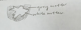
| After we located more of the structures, we made a cross sectional cut on the cerebrum, in which we were able to see the gray and white matter more distinctly. Gray matter gets its color from having more unmyelinated fibers while white matter gets its color from having high amounts of myelinated fibers. |
Structure Pin Color
Thalamus Yellow
Optic Nerve Green
Medulla Oblongata Silver
Pons White
Midbrain Blue
Corpus Callosum Red
Hypothalamus Black
 |
4) A sketch of the different structures of the inner portion of the brain.
|
5) Thalamus: Responsible for regulating sleep, consciousness, and sensory interpretation.
Optic nerve: Transfers visual information from the retina to the brain through electrical impulses.
Medulla Oblongata: Regulates breathing, heart function, and digestions.
Pons: Acts as a bridge between various parts of the nervous system.
Midbrain: Controls our vision, hearing, and sleep/wake cycles.
Corpus Callosum: Regulates our motor, sensory, and cognitive performance.
Hypothalamus: Responsible for linking the nervous system to the endocrine system through the pituitary gland.
 |
| 6) Sketch of white/gray matter |
 |
An up-close photo of the gray/white matter. The outer portion is gray matter while the inner portion is white matter.
Relate and Review:
In general, I found that the sheep dissection not only helped me understand more in depth about the anatomy and physiology of the brain but also provided me with a better picture of the functions of each part of the brain. While the lecture notes and clay brain were beneficial in providing me with a general idea of where each part of the brain is located, it was really interesting to see where each part of the brain is actually located. I also thought it was interesting to see the differences between gray and white matter. While gray matter has a gray/tan color, it has a higher concentration of unmyelinated fibers; on the other hand, white matter is a light ivory color and gets its color from the presence of high numbers of myelinated nerve fibers.
|


No comments:
Post a Comment