Thursday, April 28, 2016

Brain Dissection Reflection

Today in class, we dissected a sheep brain and located many of the brain's structures, including the cerebellum, cerebrum, midbrain, and white/grey matter. It was pretty interesting to see how many of these structures differed vastly from textbook photos/the clay brain and gave me a better understanding of the functions/structures.
The brain before removing the meninges layer. 
1) A drawing of the outer portion of the brain with anterior, posterior, cerebellum, and cerebrum labelled.

The outside of the brain after separating the meninges (3 layers of tissue that protects the brain) layer from the brain.

Structure/Location        Pin Color                 
Anterior                          White
Posterior                          Black
Cerebrum                        Yellow
Cerebellum                     Green
Brain Stem                      Silver


2/3
Functions of certain parts of the brain:
Anterior: front of the brain
Posterior: behind the brain 
Cerebellum: responsible for receiving information from the sensory systems and spinal cord; it's also responsible for regulating motor movement. 
Cerebrum: interprets information that is received from the body. 
Myelin: responsible for increasing the speed of impulses along the myelinated fiber. 


After we located more of the structures, we made a cross sectional cut on the cerebrum, in which we were able to see the gray and white matter more distinctly.  Gray matter gets its color from having more unmyelinated fibers while white matter gets its color from having high amounts of myelinated fibers.


Structure                         Pin Color 
Thalamus                        Yellow 
Optic Nerve                    Green 
Medulla Oblongata        Silver
Pons                                 White
Midbrain                          Blue
Corpus Callosum            Red
Hypothalamus                 Black

4) A sketch of the different structures of the inner portion of the brain.
5) Thalamus: Responsible for regulating sleep, consciousness, and sensory interpretation.
Optic nerve: Transfers visual information from the retina to the brain through electrical impulses.
Medulla Oblongata: Regulates breathing, heart function, and digestions.
Pons: Acts as a bridge between various parts of the nervous system.
Midbrain: Controls our vision, hearing, and sleep/wake cycles.
Corpus Callosum: Regulates our motor, sensory, and cognitive performance.
Hypothalamus: Responsible for linking the nervous system to the endocrine system through the pituitary gland.
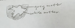
6) Sketch of white/gray matter

An up-close photo of the gray/white matter. The outer portion is gray matter while the inner portion is white matter.

Relate and Review: 
  In general, I found that the sheep dissection not only helped me understand more in depth about the anatomy and physiology of the brain but also provided me with a better picture of the functions of each part of the brain. While the lecture notes and clay brain were beneficial in providing me with a general idea of where each part of the brain is located, it was really interesting to see where each part of the brain is actually located. I also thought it was interesting to see the differences between gray and white matter. While gray matter has a gray/tan color, it has a higher concentration of unmyelinated fibers; on the other hand, white matter is a light ivory color and gets its color from the presence of high numbers of myelinated nerve fibers. 


Tuesday, April 26, 2016

Anatomy and Physiology of a Sheep Eye

 Today in class, we completed a sheep eyeball dissection, in which we used a pair of scissors, probes, tweezers, and a blade to cut through a sheep eyeball and identify the structures. It was interesting to see how each part of the eyeball play such an important role in the anatomy and physiology of the sheep; for instance, fat tissue surrounding the eye socket helps keep the eye in place when moving while the pupil regulates the amount of light entering the eye.


 Surface of Eye (Front Side)
The sclera is a white layer that forms a wall around the eyeball and helps to protect the eye from injury. We noticed that the sclera was pretty difficult to cut due to its important function.
The eyelid is responsible for shielding away dust, dirt, and other foreign particles from the eye.
The cornea is also responsible for shielding foreign particles from the eye; it acts as the eye's outermost lens and controls the entry of light in the eye. While living organisms have a clear cornea, nonliving organisms possess a cloudy cornea.


Backside of Eyeball
The fatty tissue is responsible for helping to cushion the eye when moving while the optic nerve, which is made of nerve cells uses visual information to send information from the retina to the brain.
Controls the horizontal, vertical, and rotating movements of the eye.
                                               

Inside the Eye

The retina receives light that the lens has focused and is connected to the choroid at the optic disc or the blind spot. 


The lens is responsible for changing the focal distance of the eye in order for the object to focus. This enables for an object to be formed on the retina. The suspensory ligament, which is located around the lens, is responsible for adjusting the shape of the lens to make it either more or less curved. 


The iris is the colored portion of the eye; inside, there is a pupil, which controls the amount of light that enters the eye.  The ciliary body, located beneath the iris, has many important functions, including the regulation of aqueous humor (helps maintain the shape of the eye) and the ability to have the muscle change the shape of the lens when the eyes focus on an object.


The vitreous humor has a semi-fluid consistency that fills the center cavity of the eye and is also responsible for helping to maintain the shape of the eye.  Like the cornea, the vitreous humor is sightly cloudy in a nonliving animal.  

Saturday, April 16, 2016

Clay Brain Reflection


  Today in class, we used Play-Doh to construct two models of the human brain. Through using multiple colors to represent different parts of the brain and labeling each part individually, we were able to get a better understanding of the correlation between the different parts. 


Tuesday, April 12, 2016

Hypothalamus: Necessary or Unnecessary?

   Recently, I read an article explaining a scenario in which a 24-year old woman was found to be missing a large portion of her brain, the cerebellum. Even though the cerebellum has numerous important functions and is responsible for controlling "voluntary movements and balance" and is responsible for increasing "our ability to learn specific motor actions and speak", the woman was still able to live a rather normal life with the exception of having some motor and speaking problems. It was really surprising to see that the area which the cerebellum was supposed to be had cerebrospinal fluid which allows increased cushioning to the brain and enables excess defense against various diseases. It was really surprising to see how other parts of the brain could help the body continue with most functions.
Above: Picture of a brain missing a cerebellum
Below: Actual picture of woman's brain with excess cerebrospinal fluid. 


   After reading the article, I was interested on whether or not a person could survive without a hypothalamus. I realized that unlike the cerebellum, our bodies need a hypothalamus to survive primarily because the hypothalamus is the sole part of the brain that can regulate homeostasis. This organ, located beneath the thalamus is responsible for helping the body achieve a steady rate of equilibrium or constancy. Other functions that the hypothalamus regulate include regulating blood pressure, heart rate, hunger, reflexes, thirst, bladder functions, sleep cycles, metabolism, and energy levels, making it one of the most important parts of the brain.