Wednesday, May 11, 2016

Reflexes Lab Analysis

In this lab, we tested out numerous types of reflexes, including the photo pupillary reflex, the knee jerk reflex, the blink reflex, the babe, and what's your sign reflex. Proceeding this, we tested our response times to catching a falling meterstick while texting and without it. In our notes, we learned that neurons are responsible for transmitting various messages from one area of the body to another while reflexes are involuntary actions that that the body does as a response to various stimuli. We also learned that a reflex arc is a nerve pathway. 


The Reflexes
A. The Photopupillary Reflex

Claim: As there is an increased amount of light entering the eye, there is a photopulillary reflex which causes the iris's colliery body to be stimulated. This causes it to contract. This results to the pupil decreasing in size due to the decreased amount of light entering the eye. 

Evidence: My partner covered her eye with a hand for a few minutes and after, we shined a flashlight close to an eye. The pupil's size decreased due to the decreased amount of light entering the eye. 


B. Knee Jerk Reflex

Claim: The knee jerk reflex transfers from the sensory neurons to the spinal cord and the motor neuron and finally to the knee. The knee jerk reflex is also called the monosynaptic reflex since only a single synapse in the circuit is required with the reflex. We were able to see this after repeated attempts of using the reflex hammer to find the correct part on the knee. 


Evidence: After several repeated attempts of my partner bruising my knee with a reflex hammer, my knee was able to kick out after she hit the right part of the knee. 


Reasoning: The reflex hammer causes a stretch in the thigh muscle which causes information to be sent to the spinal cord. Shortly after, the synapse is sent to the ventral horn in the spinal cord and goes back to the thigh muscle. 
C: Blind Reflex

Claim: The blind reflex is a result of the eyelid automatically closing after something rapidly comes to the eye. This was observed after both my partner and I blinked when cotton balls were thrown at us. 

Evidence: I held up a piece of saran wrap and my partner threw a cotton ball at me and I blinked. My partner also blinked when we reversed roles, showing the 
effects of the blink reflex. 

Reasoning: On average, people blink 15 times per minute or 14,400 times a day and by blinking, we are able to prevent small pieces of dust and other objects from entering our eyes.

Part 4: Babe, what's your sign

Claim: The plantar reflex occurs when the sole of the foot is stimulated using a blunt object e.g. a pen cap. When we dragged a pen from the sole of the foot to the base of the big toe, we noticed that our toes not only moved closer together but also flexed. 

Evidence: After taking off my shoes and socks, my partner took a pen with a cap and dragged it from the heel to the base of the big toe. We observed that the toes flexed and moved more closely together. 

Reasoning: This reflex is a response when the body receives a specific stimuli, showing that the plantar reflex works properly in our bodies. When there is nerve damage, there is a possibility that the toes will spread apart and upward; this is called Babinski's sign. 


Part 5: How Fast are You?

In this activity, we tested reaction time, in which our brain transferred a motor command to the arm/hand muscles. I had my partner hold the highest part of a meterstick and let it hang down while I put my hand on the bottom and was ready to catch the ruler. We documented the level at which we caught the meterstick. After three trials, we took the average time and used the table on the lab to convert the distance into the number of milliseconds. 
While my partner's time was .13 seconds, my time was .25 seconds. We perceived that the difference in time was primarily caused by the fact that my partner spends a large portion of her time playing softball; as a pitcher, she requires quick reflexes in order to avoid being hit by the ball. 

We redid the test again while texting; we realized that our times increased. For instance, my partner's time went up to .185 seconds while my time was .26 seconds; this shows that although while answering a text may appear to be important while texting, it can cause a delay in reaction time, which may cause a crash to occur. 

Class Table:
Our class's results for "How Fast are You?"

Wednesday, May 4, 2016

Learning more about the Brain

By using the Brain Map, an interactive website which allows you to locate where each part of the brain is located, I was able to learn more in depth about synapses, the frontal lobes, and the neo-cortex. 

1) What do the frontal lobes do?
The frontal lobe is responsible for a large number of functions, including controlling your behavior, remembering different tasks, and making decisions. While the left side of the frontal love is primarily language-based, the right side is responsible for other functions. 

2) What is the relationship between selective attention and learning?
Selective attention differs from learning in which it shows how selective an individual is when retaining information since it allows the memory to prioritize important information and forget about unimportant tasks. 

3) What is the last part of your brain to develop and what can you do to prevent it from deteriorating? 
The frontal lobe is the last part of your brain to develop and deteriorate. This can be stopped through exercising. 

4) What does the neo cortex do?
The neo cortex controls motor skills.

5) What is the role of the pre frontal cortex?
The pre frontal cortex is responsible for your personality and is also responsible for helping individuals to process their thoughts. 

6)What do we know about the pre frontal cortex’s relationship with multitasking?
There is a central bottleneck in the pre-frontal cortex, which acts as a mechanism that prevents people from completing multiple tasks at once. The reality is that while multitasking may seem to be an efficient way to complete numerous tasks, it negatively impacts the results of the tasks. 

7) Which part of the brain is associated with speech and language development?  Give an interesting fact about this region.
Broca's Area is responsible for speech production, language comprehension. It was interesting to see how some individuals who had defects in this region were still able to communicate with others, so other parts of the brain may have been able to complete this task. 

8) Which part of your brain is responsible for thinking the following: “Is it hot in here or is it just me?”
The Somatosensory Cortex. 

9) What does your visual cortex do for you?
The visual cortex helps to differentiate colors and different objects. 

10) State three interesting or significant facts about your occipital lobe.
The occipital lobe is responsible for processing information since it collects a large amount of visual data. It's also responsible for processing various memories.  

11) What would happen if your temporal lobes were damaged?
Because the temporal lobe is responsible for helping to hear; without it, it would be nearly impossible to listen to what other individuals were saying. 

12) What is your “fast brain” and what does it do?
This part of the brain carries out functions quickly. 

Neuron
13) State 3 things that you could do that would influence your synapses, and have a positive affect on your life and health.
Eating a healthy diet rich with nutrients and exercising are both beneficial in helping the body. Also, communicating with others (not through a screen) is an effective way to improve one's overall well-being.

14) What is the relationship between multi-sensory or multi-modal learning and your dendrites?
By continuing to remember information multiple times, it helps the brain to permanently retain the information. With various parts of the brain storing information on the same subject, it allows the brain to connect previously learned ideas with the new knowledge. 

15) How does “big picture thinking” and mnemonics affect dendrites and/or learning?
Rather than attempting to memorize solely facts, an easier way to remember ideas/concepts would be to connect the idea with something that relates to our interests or makes it easier to remember. 

16) Describe a neurotransmitter that you feel is very important.  Justify your reasoning.
Acethylcoline (ACH) is an important neurotransmitter that helps the cardiac, smooth, and skeletal muscle contract. 

Limbic System
17) What does the corpus callosum do?
It's responsible for helping you develop cognitive, motor, and sensory functions. 

18) What is the relationship between music and the corpus callosum
Music is proven to be beneficial in strengthening the two hemispheres of the brain. 

19) Why is the thalamus important?
The thalamus is responsible for regulating motor control and processing sensory information. 


Relate and Review 

Summarize what you learned from this tutorial.  Relate what you learned to your everyday life and how you can make it better.  Use at least 5 of the bolded words from the questions.  5-sentence minimum. You may use the back of this if needed.

In general, I thought that the Brain Map was pretty helpful in helping me learn about how the brain plays such a vital role in regulating cognitive/motor/sensory functions, maintaining homeostasis, learning languages, and processing information. While some parts of the brain are able to fulfill some important parts of the brain, other parts, including the hypothalamus and thalamus. In a previous blog post, I discussed why the hypothalamus's role can't be replaced be another part of the brain; with the hypothalamus being the sole part of the brain that regulates homeostasis, having a damaged hypothalamus would alter one's ability to maintain a stable body temperature. Similarly, the thalamus plays a large role in regulating the sleep/wake cycles, which plays a large role in helping us control our schedules. 

Thursday, April 28, 2016

Brain Dissection Reflection

Today in class, we dissected a sheep brain and located many of the brain's structures, including the cerebellum, cerebrum, midbrain, and white/grey matter. It was pretty interesting to see how many of these structures differed vastly from textbook photos/the clay brain and gave me a better understanding of the functions/structures.
The brain before removing the meninges layer. 
1) A drawing of the outer portion of the brain with anterior, posterior, cerebellum, and cerebrum labelled.

The outside of the brain after separating the meninges (3 layers of tissue that protects the brain) layer from the brain.

Structure/Location        Pin Color                 
Anterior                          White
Posterior                          Black
Cerebrum                        Yellow
Cerebellum                     Green
Brain Stem                      Silver


2/3
Functions of certain parts of the brain:
Anterior: front of the brain
Posterior: behind the brain 
Cerebellum: responsible for receiving information from the sensory systems and spinal cord; it's also responsible for regulating motor movement. 
Cerebrum: interprets information that is received from the body. 
Myelin: responsible for increasing the speed of impulses along the myelinated fiber. 


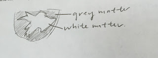
After we located more of the structures, we made a cross sectional cut on the cerebrum, in which we were able to see the gray and white matter more distinctly.  Gray matter gets its color from having more unmyelinated fibers while white matter gets its color from having high amounts of myelinated fibers.


Structure                         Pin Color 
Thalamus                        Yellow 
Optic Nerve                    Green 
Medulla Oblongata        Silver
Pons                                 White
Midbrain                          Blue
Corpus Callosum            Red
Hypothalamus                 Black

4) A sketch of the different structures of the inner portion of the brain.
5) Thalamus: Responsible for regulating sleep, consciousness, and sensory interpretation.
Optic nerve: Transfers visual information from the retina to the brain through electrical impulses.
Medulla Oblongata: Regulates breathing, heart function, and digestions.
Pons: Acts as a bridge between various parts of the nervous system.
Midbrain: Controls our vision, hearing, and sleep/wake cycles.
Corpus Callosum: Regulates our motor, sensory, and cognitive performance.
Hypothalamus: Responsible for linking the nervous system to the endocrine system through the pituitary gland.

6) Sketch of white/gray matter

An up-close photo of the gray/white matter. The outer portion is gray matter while the inner portion is white matter.

Relate and Review: 
  In general, I found that the sheep dissection not only helped me understand more in depth about the anatomy and physiology of the brain but also provided me with a better picture of the functions of each part of the brain. While the lecture notes and clay brain were beneficial in providing me with a general idea of where each part of the brain is located, it was really interesting to see where each part of the brain is actually located. I also thought it was interesting to see the differences between gray and white matter. While gray matter has a gray/tan color, it has a higher concentration of unmyelinated fibers; on the other hand, white matter is a light ivory color and gets its color from the presence of high numbers of myelinated nerve fibers. 


Tuesday, April 26, 2016

Anatomy and Physiology of a Sheep Eye

 Today in class, we completed a sheep eyeball dissection, in which we used a pair of scissors, probes, tweezers, and a blade to cut through a sheep eyeball and identify the structures. It was interesting to see how each part of the eyeball play such an important role in the anatomy and physiology of the sheep; for instance, fat tissue surrounding the eye socket helps keep the eye in place when moving while the pupil regulates the amount of light entering the eye.


 Surface of Eye (Front Side)
The sclera is a white layer that forms a wall around the eyeball and helps to protect the eye from injury. We noticed that the sclera was pretty difficult to cut due to its important function.
The eyelid is responsible for shielding away dust, dirt, and other foreign particles from the eye.
The cornea is also responsible for shielding foreign particles from the eye; it acts as the eye's outermost lens and controls the entry of light in the eye. While living organisms have a clear cornea, nonliving organisms possess a cloudy cornea.


Backside of Eyeball
The fatty tissue is responsible for helping to cushion the eye when moving while the optic nerve, which is made of nerve cells uses visual information to send information from the retina to the brain.
Controls the horizontal, vertical, and rotating movements of the eye.
                                               

Inside the Eye

The retina receives light that the lens has focused and is connected to the choroid at the optic disc or the blind spot. 


The lens is responsible for changing the focal distance of the eye in order for the object to focus. This enables for an object to be formed on the retina. The suspensory ligament, which is located around the lens, is responsible for adjusting the shape of the lens to make it either more or less curved. 


The iris is the colored portion of the eye; inside, there is a pupil, which controls the amount of light that enters the eye.  The ciliary body, located beneath the iris, has many important functions, including the regulation of aqueous humor (helps maintain the shape of the eye) and the ability to have the muscle change the shape of the lens when the eyes focus on an object.


The vitreous humor has a semi-fluid consistency that fills the center cavity of the eye and is also responsible for helping to maintain the shape of the eye.  Like the cornea, the vitreous humor is sightly cloudy in a nonliving animal.  

Saturday, April 16, 2016

Clay Brain Reflection


  Today in class, we used Play-Doh to construct two models of the human brain. Through using multiple colors to represent different parts of the brain and labeling each part individually, we were able to get a better understanding of the correlation between the different parts. 


Tuesday, April 12, 2016

Hypothalamus: Necessary or Unnecessary?

   Recently, I read an article explaining a scenario in which a 24-year old woman was found to be missing a large portion of her brain, the cerebellum. Even though the cerebellum has numerous important functions and is responsible for controlling "voluntary movements and balance" and is responsible for increasing "our ability to learn specific motor actions and speak", the woman was still able to live a rather normal life with the exception of having some motor and speaking problems. It was really surprising to see that the area which the cerebellum was supposed to be had cerebrospinal fluid which allows increased cushioning to the brain and enables excess defense against various diseases. It was really surprising to see how other parts of the brain could help the body continue with most functions.
Above: Picture of a brain missing a cerebellum
Below: Actual picture of woman's brain with excess cerebrospinal fluid. 


   After reading the article, I was interested on whether or not a person could survive without a hypothalamus. I realized that unlike the cerebellum, our bodies need a hypothalamus to survive primarily because the hypothalamus is the sole part of the brain that can regulate homeostasis. This organ, located beneath the thalamus is responsible for helping the body achieve a steady rate of equilibrium or constancy. Other functions that the hypothalamus regulate include regulating blood pressure, heart rate, hunger, reflexes, thirst, bladder functions, sleep cycles, metabolism, and energy levels, making it one of the most important parts of the brain. 

Thursday, March 24, 2016

Unit 7 Blog Reflection

Wednesday, March 23, 2016

Caffeine Post

 Today in class, we created an ad demonstrating the effects of caffeine. Even though caffeine may be viewed as a relatively safe substance since it's not only legal but can also be purchased at a coffee shop located on nearly every street corner by even young children, the reality is that caffeine can easily become addictive and has rather negative side effects. Even though caffeine has the ability to stimulate one's mind to allow one to cram for a test or finish a homework assignment, the drug has a tendency to cause insomnia, nervousness, dehydration, and irritability.
 One thing that concerns me with caffeinated beverages, specifically energy drinks, is the amount of caffeine each one has. After doing some research, I found a website stating that a 16 oz. can of Monster has about 160 mg. of caffeine, which is "more caffeine than a double espresso". It was really surprising to see that a 16 ounce can of an energy drink has over half of the maximum recommended dose of caffeine daily. Since most consumers are probably not aware of the possible side effects that caffeine has, I feel that all bottled and canned drinks should have a small label warning consumers of the possible side effects that may occur with caffeine consumption.

Monday, March 21, 2016

Chicken Dissection


 On Thursday, we spent the class period dissecting a chicken carcass using a scalpel, a probe, and tweezers while labeling the various muscle structures and comparing the chicken's physical and functional differences to the human's. Because we went over the majority of the muscles previously in the lecture, it was easy to locate the general area of each muscle on the chicken's body.
 Muscles, bones, and tissues all play important functions in our bodies; for instance, our bones, which are composed of dense connective tissue, are responsible for supporting the body, protecting the body's soft organs, and forming blood cells. Tendons are flexible chords of fibrous tissue that is responsible for attaching a muscle to a bone.  For instance, the biceps are joined together by a tendon at the radius and ulna. Muscles are responsible for stabilizing joints, heat generation, maintaining the body's posture, and moving the body's bones and fluids.

 Overall, we noticed that the chicken had many functional similarities but had vastly different physical similarities. Because the chicken that we dissected was bred primarily for meat, we noticed that its body was extremely unbalanced and often toppled over, with parts of their bodies (e.g. the pectoralis minor and major) much larger in proportion than other parts of their body. On the other hand, most humans seem to have relatively proportional bodies. Another difference that we found was that the upper limbs of humans are arms while chickens possess wings as their upper limbs. While chickens and humans possess structural differences, both use their upper limbs for the same function. One similarity that we found between humans and chickens is that both possess epithelial, muscle, and connective tissue.

The pectoralis major is the breast muscle that runs from the shoulder to the humerus

The pectoralis minor lifts the wings dorsally which allows the chicken to recover when flying.

In chickens, the trapezius runs perpendicular 
from the backbone to the shoulder of the bird 
and is responsible for pulling the shoulders back.
In humans, the trapezius is split in 2 parts. 

The deltoid helps the raise the upper arm/wing.
The biceps is responsible for bending the arm/wing.
The triceps are responsible for straightening the arm/wing.

This is the chicken's largest muscle on the posterior side of the chicken's lower wing.
In humans, this muscle is responsible for flexing the hand.
This is the chicken's largest muscle on the superior side of the lower wing.
In humans,this muscle is responsible for pulling the hand back.

The sartorius is responsible for flexing the thighs and crossing the legs.


The iliubialis covers the lateral side of the chicken's thigh. In humans, it runs from the dorsal and lateral sides of the pelvis to the lateral side of the knee.


The biceps femoris lies medial to the inferior section of the iliotibialis and is responsible for flexing the leg.


The semimembranosus is inferior and medial to the biceps femoris and is responsible for extending the thigh.


The semitendinosus is anterior and medial to the semimembranosus and extends the thigh.


The quadriceps femoris lies on the inside of the thigh medial to the sartorius and is responsible for flexing the thigh and extending the lower leg. 


The gastrocnemius is the primary muscle of the drumstick. In both humans and chickens, the gastrocnemius is attached to the Achilles tendon/

Whole chicken before the dissection


An example of some of the chicken's tendons